Session 6
12 April 2022
4:30 - 6:00pm SGT (UTC+8)
5:30 - 7:00pm JST
6:30-8:00pm AEST
12 April 2022
4:30 - 6:00pm SGT (UTC+8)
5:30 - 7:00pm JST
6:30-8:00pm AEST
Abstract
The extra-cellular matrix (ECM) is a source of biochemical and biomechanical influences that direct cell proliferation and fate. However, the molecular mechanisms that regulate the interplay between these influences and tissue homeostasis are still relatively ill-defined. While cancer cells forming tumours of epithelial origin do not frequently synthesise substantial amounts of ECM proteins, they strongly influence genetically normal cells within the tumour microenvironment to do so, resulting in a dense ECM, configured for tumour-promotion. We have shown that cutaneous squamous cell carcinomas produce CRELD2, which we have previously shown to recruit and educate cancer-associated fibroblasts, which then produce a dense, collagen-rich ECM that promotes tumour progression. CRELD2 production is regulated by signalling through the RHO-ROCK pathway and PERK-regulated transcription via the ATF4, providing exploitable opportunities for targeting it. Conversely, the RHO-ROCK pathway regulates the re-establishment of the dermal ECM during skin wound healing, and we have established a new way to take advantage of this process to accelerate healing in a model of diabetic wounds. Exploiting effector pathways downstream of ROCK may therefore be useful for new mechanotherapy approaches to deal with conditions as diverse as cancer and chronic wounds.
The extra-cellular matrix (ECM) is a source of biochemical and biomechanical influences that direct cell proliferation and fate. However, the molecular mechanisms that regulate the interplay between these influences and tissue homeostasis are still relatively ill-defined. While cancer cells forming tumours of epithelial origin do not frequently synthesise substantial amounts of ECM proteins, they strongly influence genetically normal cells within the tumour microenvironment to do so, resulting in a dense ECM, configured for tumour-promotion. We have shown that cutaneous squamous cell carcinomas produce CRELD2, which we have previously shown to recruit and educate cancer-associated fibroblasts, which then produce a dense, collagen-rich ECM that promotes tumour progression. CRELD2 production is regulated by signalling through the RHO-ROCK pathway and PERK-regulated transcription via the ATF4, providing exploitable opportunities for targeting it. Conversely, the RHO-ROCK pathway regulates the re-establishment of the dermal ECM during skin wound healing, and we have established a new way to take advantage of this process to accelerate healing in a model of diabetic wounds. Exploiting effector pathways downstream of ROCK may therefore be useful for new mechanotherapy approaches to deal with conditions as diverse as cancer and chronic wounds.
Bio
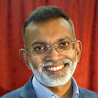
Michael Samuel is a cell biologist with an interest in understanding the cellular and non-cellular microenvironments of epithelial cancers and how they are established to promote tumour growth. He heads the Tumour Microenvironment Laboratory at the Centre for Cancer Biology and is Professor of Matrix Biology at the University of South Australia, Adelaide. He obtained his PhD from the University of Melbourne in 2004 and then joined the laboratory of Prof. Mike Olson at the Beatson Institute for Cancer Research, Glasgow, with whom he identified the role of the Rho-ROCK signalling pathway in regulating mechanical properties of the extracellular matrix. He returned to Australia in 2012 to join the Centre for Cancer Biology as a Principal Investigator. His laboratory investigates the mechanisms by which cancer cells co-opt genetically normal cells within the microenvironment to promote tumour growth and metastasis, employing inter-disciplinary approaches spanning cell and tissue biology, and the materials sciences. He has been awarded an Australian Research Council Future Fellowship, an Emerging Leader Award by the Australia and New Zealand Society for Cell and Developmental Biology and the Barry Preston Award by the Matrix Biology Society of Australia and New Zealand.
Michael Samuel is a cell biologist with an interest in understanding the cellular and non-cellular microenvironments of epithelial cancers and how they are established to promote tumour growth. He heads the Tumour Microenvironment Laboratory at the Centre for Cancer Biology and is Professor of Matrix Biology at the University of South Australia, Adelaide. He obtained his PhD from the University of Melbourne in 2004 and then joined the laboratory of Prof. Mike Olson at the Beatson Institute for Cancer Research, Glasgow, with whom he identified the role of the Rho-ROCK signalling pathway in regulating mechanical properties of the extracellular matrix. He returned to Australia in 2012 to join the Centre for Cancer Biology as a Principal Investigator. His laboratory investigates the mechanisms by which cancer cells co-opt genetically normal cells within the microenvironment to promote tumour growth and metastasis, employing inter-disciplinary approaches spanning cell and tissue biology, and the materials sciences. He has been awarded an Australian Research Council Future Fellowship, an Emerging Leader Award by the Australia and New Zealand Society for Cell and Developmental Biology and the Barry Preston Award by the Matrix Biology Society of Australia and New Zealand.
Abstract
Methods for culturing human embryonic stem cells (hESC) enable their use in generation of various hESC-derived cells after in vitro differentiation. Such cells could be used in regenerative medicine to treat damaged tissues such as heart infarction that normally cannot renew, blindness due to loss of photoreceptors or type 1 diabetes due to death of islet cells. However, such cells may contain tumorigenic cells, or they may not contain cell types that are not suitable for the purpose and as therapeutic cells. We have developed methods to differentiate hESCs under completely defined and xenogen-free conditions and are reproducible differentiation methods. The pluripotent cells are cultured on various human basement membrane laminins that are important during embryogenesis where they underlie or surround all organized cells in the body. We have shown that e.g. highly specific laminins drive differentiation of hESCs to cardiac muscle cells, retina laminins drive differentiation to RPEs and photoreceptor lineage, or we can expand keratinocyte stem cells in large amounts for treatment of burn wounds. Experiments with in vitro differentiated cells have shown the importance of using biologically relevant laminins in the of hESCs differentiated on biologically relevant laminins that are normal components of stem cell differentiation during embryogenesis and organogenesis.
By culturing pluripotent hESCs on specific cell type specific recombinant laminins, we can drive differentiation the cells to various lineages, such as cardiomyocytes and progenitors, retinal RPEs and photoreceptor progenitors and for the first time we have shown it to be possible to culture human skin keratinocytes instead of using xenografts with human keratinocytes and mouse fibroblast feeder cells. Cardiomyocyte progenitors injected into the heart infarction region of swines form human cardiomyocyte fiber bundles and exhibit improved heart function. Also, photoreceptor surrounding retina laminins specifically enable generation of progenitor cells that express typical photoreceptor markers and improve heart function. Differentiation of the pluripotent stem cells on certain laminins turns of the expression of tumorigentic cells and we do not see signs of teratoma formation. Together with coworkers Alvin Chua and others at Duke-NUS and SGH, we are culturing autologous keratinocyte stem cells for treatment of burn wounds.
Bio
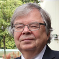
Karl Tryggvason, MD, PhD, is Professor at Duke-NUS Medical School, Singapore and Duke University, North Carolina, as well as Professor Emeritus at the Karolinska Institute. His group has cloned and produced most human BM proteins and clarified genetic causes of many BM-associated diseases, such as Alport and congenital nephrotic syndromes, junctional epidermolysis bullosa and congenital muscular dystrophy, as well as studied matrix metalloproteinases, including the discovery and crystal structure of MMP-2. His group has produced most laminins as recombinant human proteins and currently the group focuses on how different laminin isoforms influence cell identity, growth and stem cell differentiation. Tryggvason has published over 400 research articles. He is a member of the Finnish Academy of Sciences and the Swedish Royal Academy of Sciences, and he has served for 18 years as a member of the Nobel Assembly and Committee for Physiology or Medicine at the Karolinska Institute. He has received multiple international awards. He is co-founder of BioLamina AB, Stockholm, that produces laminins for cell biology and stem cell derived cells aimed for regenerative medicine.
Karl Tryggvason, MD, PhD, is Professor at Duke-NUS Medical School, Singapore and Duke University, North Carolina, as well as Professor Emeritus at the Karolinska Institute. His group has cloned and produced most human BM proteins and clarified genetic causes of many BM-associated diseases, such as Alport and congenital nephrotic syndromes, junctional epidermolysis bullosa and congenital muscular dystrophy, as well as studied matrix metalloproteinases, including the discovery and crystal structure of MMP-2. His group has produced most laminins as recombinant human proteins and currently the group focuses on how different laminin isoforms influence cell identity, growth and stem cell differentiation. Tryggvason has published over 400 research articles. He is a member of the Finnish Academy of Sciences and the Swedish Royal Academy of Sciences, and he has served for 18 years as a member of the Nobel Assembly and Committee for Physiology or Medicine at the Karolinska Institute. He has received multiple international awards. He is co-founder of BioLamina AB, Stockholm, that produces laminins for cell biology and stem cell derived cells aimed for regenerative medicine.
CHAIRS
Nikolas HAASS, The University of Queensland Diamantina Institute, Australia
Hironobu FUJIWARA, RIKEN, Japan
Nikolas HAASS, The University of Queensland Diamantina Institute, Australia
Hironobu FUJIWARA, RIKEN, Japan


